Download de presentatie
De presentatie wordt gedownload. Even geduld aub

1
Mark Winkens Tweesteden Ziekenhuis, Tilburg
Cardiovasculair risicomanagement anno 2011 CT angiografie Mark Winkens Tweesteden Ziekenhuis, Tilburg

2
Casus Vrouw 1956 VG: Hypercholesterolemie, Hypertensie
RF: Familiair belast Rx: Simvastatine

3
met verhoogd risicoprofiel en licht afwijkend ecg
Casus Presentatie EHH ivm zowel typische als atypische thoracale klachten Lichamelijk onderzoek: RR 180/90 L=R, p 90 bpm, verder g.b. Conclusie: deels typische deels atypische thoracale pijn bij 53 jarige vrouw met verhoogd risicoprofiel en licht afwijkend ecg lab: trop T < 0.01 Grace score 6 maanden dood of MI: 7% Plan: direct Cardiale CT ter rule out coronairlijden

4
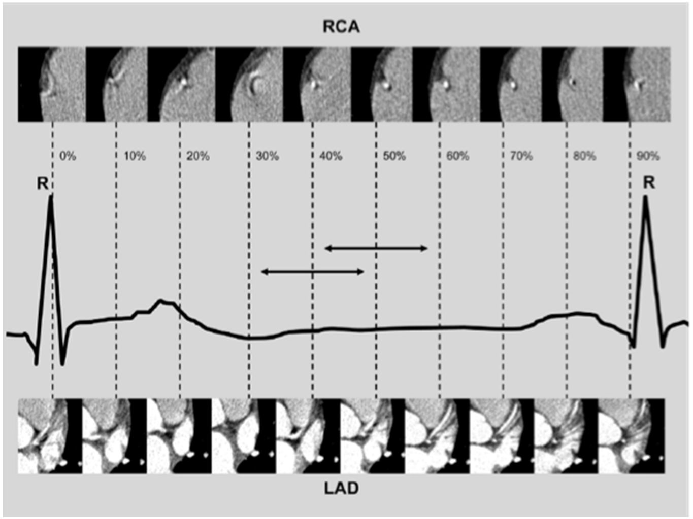
At least 180 degrees of data are required for image reconstruction
CT data collection rotatie tijd 270 ms!! X-ray tube Detector array At least 180 degrees of data are required for image reconstruction

6
Multislice scanners (MSCT), multi detector (MDCT)
combi met PET slice Philips, Toshiba, Siemens, GE Dual source scanners (Siemens) Toekomst: Dual layer scanners Dual energy scanners Spectral CCT
 Toekomst: Dual layer scanners. Dual energy scanners. Spectral CCT.")
7
Iterative reconstruction methods
Dose exposure Iterative reconstruction methods 0,6-2 mSv Just to come back to reality I want to show you this slide. The mean dose exposure for a chest X-ray is 0.2 mSV. The natural yearly dose exposure in central europe is 2mSv.The dose exposure for cardiac catherization is 5 -8 mSV depending on the amount of positions the cardiologist needs. The dose exposure for a cardiac CT with helical scan mode is 15 mSV. This is still a lot and so we fully agree that it is very important to seriously check the indication of any kind of CT. high end Cardiac - CTa 1-3 mSv Courtesy: Dr.O. Klass/Dr.M. Hoffmann BU-CT, DACH, Gerold Krüger, Hamburg 2

8
Current status in coronary artery imaging (64 slice)
tekst CCT; indications, applications, limitations, and traing requirements EHJ (2008) 29,
 29,")
10
Medmar, Istanbul, Turkey
Images Courtesy of: Medmar, Istanbul, Turkey Current status : Low dose higher HR with arrhythmia 128x0.625 RT 0.27s Scan Time 5s Scan Length 12 cm 120kV, 200mAs 2.8mSv 3 3

11
Excellent robust image quality
RT 0.27s S&S no tolerance Sharp kernel XCC Scan Length 28 cm 100kV,150mAs

12
20778 Chest Pain Assessment 1463 images reconstructed in 90 s! “ Triple rule out” Acute chest pain, aortic dissection 46 yrs Brilliance iCT 100 kVp Step & Shoot Complete Adaptive Collimation iDose4 3.5 mSv Normal ECG, enzyme test negative…so what is the issue in this patient with acute chestpain : PE, Dissection or Coronary issues ? S&S + iDose wipes the dose issue of the table ( dose was one of the main reasons why clinicians did not start immediately with this kind of protocol) S&S works with HR up to 75, Flash needs a max of 60bpm…and Tosh has problems over 65bpm…so no real threat of the competitors here. GE is poor in coverage and dose and Tres…. Study shows a dissection of the Aorta as reason for the chestpain. No PE, no coronary lesions. One study shows it all !!! only 3.5mSv…. 12
 S&S works with HR up to 75, Flash needs a max of 60bpm…and Tosh has problems over 65bpm…so no real threat of the competitors here. GE is poor in coverage and dose and Tres…. Study shows a dissection of the Aorta as reason for the chestpain. No PE, no coronary lesions. One study shows it all !!! only 3.5mSv…. 12.")
13
Fusion of Nuclear. Med and CT: PET and SPECT CT

14
Cardiac CT the (non invasive) athero burden read out of the future in your practice and research??
 athero burden read out of the future in your practice and research")
15
Accuracy of Plaque detection
Detection rate MSCT vs IVUS: Soft Plaque: 83% (54/65) Mixed Plaque: 94% (50/53) Calcified Plaque: 95% (41/43) Leber et. al. Accuracy of 64-slice computed tomography to classify and quantify plaque volumes in the proximal coronary system: a comparative study using intravascular ultrasound. J Am Coll Cardiol, 47(3), , 2006. Correlation IVUS vs CT quant: r And also these images from the iCT show an improvement of the accuracy of plaque detection. According to Leber et al. there is a good correlation between IVUS and MSCT even at a 64 row scanner. They found that CT detected 83% of softplaques, 94% of mixed plaques and 95% of calcified plaques compared to IVUS. Sample CT coronary angiogram iCT Leber et. al. Accuracy of 64-slice computed tomography to classify and quantify plaque volumes in the proximal coronary system: a comparative study using intravascular ultrasound. J Am Coll Cardiol, 47(3), , 2006.
 Mixed Plaque: 94% (50/53) Calcified Plaque: 95% (41/43) Leber et. al. Accuracy of 64-slice computed tomography to classify and quantify plaque volumes in the proximal coronary system: a comparative study using intravascular ultrasound. J Am Coll Cardiol, 47(3), , Correlation IVUS vs CT quant: r And also these images from the iCT show an improvement of the accuracy of plaque detection. According to Leber et al. there is a good correlation between IVUS and MSCT even at a 64 row scanner. They found that CT detected 83% of softplaques, 94% of mixed plaques and 95% of calcified plaques compared to IVUS. Sample CT coronary angiogram. iCT. Leber et. al. Accuracy of 64-slice computed tomography to classify and quantify plaque volumes in the proximal coronary system: a comparative study using intravascular ultrasound. J Am Coll Cardiol, 47(3), ,")
16
Klass et al.; Int J Cardiovasc Imaging 2010

17
Plaque Imaging Softplaque 2005 rule out indication
For the next next scanner generation we are now equiped with a new software for plaque evaluation and volumetry. This might be very interesting for monitoring the total volume of soft plaque burden for e.g. to monitor a statin therapy. This srceen shot shows a single softplaque of the LAD. The histogramm below shows distribution of plaque components in terms of calcified or soft plaque tissue.

18
-after 4 years of statin therapy-
Plaque monitoring Softplaque 2009 -after 4 years of statin therapy- For the next next scanner generation we are now equiped with a new software for plaque evaluation and volumetry. This might be very interesting for monitoring the total volume of soft plaque burden for e.g. to monitor a statin therapy. This srceen shot shows a single softplaque of the LAD. The histogramm below shows distribution of plaque components in terms of calcified or soft plaque tissue.

19
The High risk plaque JACC july 2009

20
The Napkin-Ring Sign: the high risk coronary plaque?
JACC cv imaging Vol 3 No : Maurovich, Hoffman, Virmani et al.

21
JACC vol. 58 no.19 nov :

22
5924 Improving Positive Predictive Value Anatomy + Perfusion : One Stop background radiation Recurrent chest pain, old LAD stent 42 yrs Brilliance iCT 100 kVp Step & Shoot iDose4 3.0 mSv Myocardial Defect Assessment Physicians are looking for ways to improve pos. predictive value. Many C-CTA’s show a sign calc. plaque, which makes it impossible to rate it… So you either can send the patient to the CATH ( which might be not needed…) or add an additional test for physiology…stress/rest MR or SPECT. But this involves more modalities……it would be great if CT could do it all…. Large multi center trial started in USA to proof CT rest/stress static perfusion is as good as a SPECT or MR…. Case shows a defect in rest ( so an infarct), but more important to stress is that Intellispace Portal is ready for the ( near) future. tekst 22
 or add an additional test for physiology…stress/rest MR or SPECT. But this involves more modalities……it would be great if CT could do it all…. Large multi center trial started in USA to proof CT rest/stress static perfusion is as good as a SPECT or MR…. Case shows a defect in rest ( so an infarct), but more important to stress is that Intellispace Portal is ready for the ( near) future. tekst. 22.")
Verwante presentaties


>")


>")


