Download de presentatie
De presentatie wordt gedownload. Even geduld aub

1
ESC echo examen TTE Indeling: Fysiologie Formules Normaalwaardes
Guidelines

2
Fysiologie Echogolven: backscatter, refractie en transmissie
Backscatter produceert het meeste van het ultrasound beeld Refractie en transmissie komen niet terug naar de transducer

3
Fysiologie formules V = f x wavelength
Snelheid ultrasound soft tissue 1540m/s Gemakkelijke formule: 1.54 / Mhz = wavelength in mm Snelheid geluid in bot hoger dan bloed, soft tissue, lucht L (length of the nearfield) = r 2 / wavelength Hoe hoger de frequentie, hoe langer de nearfield Doppler shift = 2 x f 0 x v / c cos hoek
 = r 2 / wavelength. Hoe hoger de frequentie, hoe langer de nearfield. Doppler shift = 2 x f 0 x v / c cos hoek.")
4
Fysiologie Pulse duration or length (mm) PRF Duty factor (N 0.1%)
Physical length that pulse occupes Affected by source of ultrasound PRF Rate at which pulses are emitted from transducer 77.000/depth in cm Duty factor (N 0.1%) Fraction of time that transducer is emitting ultrasound Pulse duration / dead time Impedance Rayls: density in kg/m3 x speed of sound in m/s Differences in impedance determine the ratio of transmitted versus reflected sound Hoe hoger frequentie: hoe hoger de resolutie, maar hoe minder het doordringingsvermogen
 Fraction of time that transducer is emitting ultrasound. Pulse duration / dead time. Impedance. Rayls: density in kg/m3 x speed of sound in m/s. Differences in impedance determine the ratio of transmitted versus reflected sound. Hoe hoger frequentie: hoe hoger de resolutie, maar hoe minder het doordringingsvermogen.")
5
Resolutie Spatial resolutation Temporal resolutie Axial resolution (Z)
Pulse length, transducer frequency Lateral resolution (X, Y) Beam width, depth, gain Temporal resolutie Synoniem = frame rate Depth, sweep angle, line density, PRF
 Beam width, depth, gain. Temporal resolutie. Synoniem = frame rate. Depth, sweep angle, line density, PRF.")
6
Belangrijke formules PISA Continuity equation Bernouilli Flow en CO
PHT

7
PISA Principe = PISA flow gelijk aan MR flow ERO = flow rate / MR Vmax
2 pi r2 x Vn (Q in cc / sec) = ERO x MR Vmax ERO = flow rate / MR Vmax MR velocity in cm / s ERV = ERO x VTI MR
 = ERO x MR Vmax. ERO = flow rate / MR Vmax. MR velocity in cm / s. ERV = ERO x VTI MR.")
8
Bernouilli Snelheid is een schatting van de drukgradient over een klep, naar aanleiding van de eerste wet van behoud van energie (Newton) Vereenvoudigde vergelijking Drukgradient = 4 x v 2 Niet meegenomen: deceleratie na klep en viscositeit bloed Wel voor berekening: Stenotische klep (AS) PH met TI signaal + RAP (5/10/15) LVOT contour en VTI Snelheid VSD: LVsp – PGjet = RVsp = sPAP Een lage snelheid over VSD betekent een hoge sPAP Eind diastolische snelheid PI jet: drukberekening is edRVdruk + RAP Schatting PVR MR (Dp/Dt): LV pressure increase early systolic: EH mmHg/sec = 32/tijdsinterval in sec Niet te gebruiken bij dubbele klepvitiae, bij aorta ascendens van minder dan 30mm (pressure recovery), bij V1 > 1 m/s
 PH met TI signaal + RAP (5/10/15) LVOT contour en VTI. Snelheid VSD: LVsp – PGjet = RVsp = sPAP. Een lage snelheid over VSD betekent een hoge sPAP. Eind diastolische snelheid PI jet: drukberekening is edRVdruk + RAP. Schatting PVR. MR (Dp/Dt): LV pressure increase early systolic: EH mmHg/sec = 32/tijdsinterval in sec. Niet te gebruiken bij dubbele klepvitiae, bij aorta ascendens van minder dan 30mm (pressure recovery), bij V1 > 1 m/s.")
9
Continuity equation Tweede wet van Newton: law of conservation of mass
Mass cannot be destroyed, flow rates are the same at different locations in a flow stream Flow rate op punt 1 is gelijk aan punt 2 A1 x V1 = A2 x V2 LVOT area x LVOT VTI = AVA x aortic VTI AVA = LVOT area x LVOT VTI/aortic VTI

10
Flow en CO Flow rate = CSA x flow velocity (Vmax)
SV = CSA x VTI = pi 2 r x VTI Assuming a circular shape Ook voor berekening RV / R fractie / Qp : Qs (intracardiac shunt) CO = SV x HF Fick Less than transaortic flow (Fick + RV)
 CO = SV x HF. Fick. Less than transaortic flow (Fick + RV)")
11
Doppler Aliasing Doppler tissue imaging
Nyquist limit is highest obtainable velocity When frequency is higher than Nyquist Nyquist limit = PRF/2 Doppler tissue imaging Filter out low amplitudes and high Doppler shifts MPI CW 4/5CH: IVCT (systole)/IVRT (diastole) / ET
/IVRT (diastole) / ET.")
12
Hemodynamiek Flow: laminair/turbulent Vroege sluiting MK: AoI
B-hump: vertraagde sluiting MK, verhoogde einddiastolische LV druk Vroegsystolisch naar beneden bewegen IVS: LBTB Afplatting IVS: systolisch (drukbelasting RV), diastolisch (volumebelasting RV) IVC dilatatie: verhoogde RA druk
, diastolisch (volumebelasting RV) IVC dilatatie: verhoogde RA druk.")
13
Artefacten Side lobes Reverberaties Shadowing Near-field clutter
Posterieure mitralisklepannulus Reverberaties Posterieure pericard Shadowing Kunstkleppen Near-field clutter Lijkt op LV thrombus

14
Normaalwaardes Bij mannen van volwassen leeftijd

15
LV FS = (LVEDD-LVESD)/LVEDD x 100%
LV massa = 0.8 x 1.04 x (IVS+LVPW+LVEDD) g LVMI = LV massa/BSA Myocardial volume = LV volume-blood pool volume
 g. LVMI = LV massa/BSA. Myocardial volume = LV volume-blood pool volume.")
16
LV Septum < 10mm Posterior wand <10mm LVEDD <59mm
LVEDV <155ml LVESV <58ml EF >55% FS >25% LV massa <224g LVMI <115g/m2 WMSI <1.5 dP/dT >1200mmHg/s LV volume d+s (Simpson), SV Lateral annulus motion (N>16mm), E-point septal separation (EPSS, N<6mm), aortic valve closure in M-mode
, SV. Lateral annulus motion (N>16mm), E-point septal separation (EPSS, N<6mm), aortic valve closure in M-mode.")
17
RWBS Visueel, WMSI (stroomgebieden) Nonischemisch: geleiding (LBTB, PM, PVC, WPW), constrictie, tardokinesis/vroege relaxatie Kwantitatief Doppler Meting flow (CSA x Vmax) en SV (CAA x VTI) MPI (IVCT + IVRT / ET of TST-ET/ET) (N<0.4) dP/dT op MI signaal, drukverschil 32mmHg/tijd, N>900mmHg/sec Strain en strain rate via Doppler (TDI) of speckle tracking
 en SV (CAA x VTI) MPI (IVCT + IVRT / ET of TST-ET/ET) (N<0.4) dP/dT op MI signaal, drukverschil 32mmHg/tijd, N>900mmHg/sec. Strain en strain rate via Doppler (TDI) of speckle tracking.")
19
RV RV mediolateraal midventriculair d 35mm RV lengte d <79mm
RV lengte s <63mm RV oppervlakte <28cm2 RV dikte < 5mm TAPSE >16mm RVOT <2.3mm

20
Diastologie Diastole bestaat uit: IVRT, early filling (reservoir), diastasis, atrial systole (pomp) TTE: E/A (LA-LV gradient, N>1) Deceleratietijd (compliance, prognosis MI, N ms) E’ (ventricular relaxation, LV filling pressure in diastolic dysfunction, lateraal hoger dan septaa, N>12cm/sl) E/E’ (LV filling pressure=LVEDP, N 5-10) IVRT (LA pressure, early active LV relaxation, N 80ms) S/D/A pulmonaalvenen (N S>D>A), Ar-A (LV filling pressure, N 0) LA volume (chronicity, prognosis, N<29ml/m2) Vp (early diastolic relaxation, estimation PCWP) , N>50cm/s) LV functie sPAP Valsalva (differentiating stages, in pseudonormal stage E decreases, A increases) LVEDP < 16mmHg, PCWP <12mmHg Maat voor relaxatie (invasief): Tau
 Deceleratietijd (compliance, prognosis MI, N ms) E’ (ventricular relaxation, LV filling pressure in diastolic dysfunction, lateraal hoger dan septaa, N>12cm/sl) E/E’ (LV filling pressure=LVEDP, N 5-10) IVRT (LA pressure, early active LV relaxation, N 80ms) S/D/A pulmonaalvenen (N S>D>A), Ar-A (LV filling pressure, N 0) LA volume (chronicity, prognosis, N<29ml/m2) Vp (early diastolic relaxation, estimation PCWP) , N>50cm/s) LV functie. sPAP. Valsalva (differentiating stages, in pseudonormal stage E decreases, A increases) LVEDP < 16mmHg, PCWP <12mmHg. Maat voor relaxatie (invasief): Tau.")
21
Diastologie Zie volgende tabel + E 72 cm/s A 40 cm/s
Verhoogde LA druk bij: IVRT < 70ms E/A > 2 Deceleratietijd <150ms S/D < 1 Toegenomen AR-duur en amplitude E/E’ > 15 LA dilatatie

22
EAE

23
Gradering 1 2 3 4 Impaired relaxation Pseudonormalisation
Delayed LV early diastolic active relaxation, normal LA pressure IVRT lengthens 2 Pseudonormalisation Mildly elevated LA pressure, low LA-LV gradient, reduced LV suction force High filling pressure combined with impaired relaxation E wave reduced, IVRT reduced 3 Restrictive filling Noncompliant LV chamber, increased stiffness ,elevated LA pressure, high opening LA-LV pressure gradient, failing LA contractility, responds positively to preload reduction (Valsalva, diurese) Ar-A>30ms 4 Restrictive filling, irreversible Inflow by pushing blood
 Ar-A>30ms. 4. Restrictive filling, irreversible. Inflow by pushing blood.")
24
Constrictief vs restrictief
Restrictief (diastolische dysfunctie graad 3, amyloidose) LA > LVF N E/A > 1.5 (geen verandering op preloadvermindering) DT < 160ms Es’ < 8cm/sec PH, TI>MI, inspiratoire diastolische leverveneflowomkering Constrictief Valsalva: >25% verandering E IVRT variatie met ademhaling
 LA > LVF N. E/A > 1.5 (geen verandering op preloadvermindering) DT < 160ms. Es’ < 8cm/sec. PH, TI>MI, inspiratoire diastolische leverveneflowomkering. Constrictief. Valsalva: >25% verandering E. IVRT variatie met ademhaling.")
25
Atria LA <30mm RA <45mm LA <20mm2 LA volume minder dan 58ml
LA-volume/BSA < 28ml/m2 Flowsnelheid bij TTE N > 64cm/sec Verhoogde kans op thrombusvorming bij >30cm/sec Schatting RA-druk: 5 RA N 10 Vmax TI 2.6-4m/s VCI gedilateerd 15 Severe TI Geen respiratoire variatie VCI

26
Longvenen S1: atriale relaxatie S2: inflow door RV contractie
S-flowsnelheid cm/sec D-flowsnelheid cm/sec

27
Aorta Boog <30mm Descendens <30mm Abdominalis <30mm

28
Schatting sPAP sPAP = 4x Vmax TI + RA druk
Pulmonale acceleratietijd >120ms

29
MI Jetgrootte <4cm2 of <20% van LA oppervlakte = geringe MI Flow-convergentie zone van <0.9cm = geringe MI Regurgitatiefractie <30% = geringe MI Morfologie: indeling volgens Carpentier (normale bewegelijkheid I, verhoogde bewegelijkheid II, verminderde bewegelijkheid III) Mitralisklepannulusdiameter (N <35 mm) Likelihood repair: calcificatie, tenting area >2.5cm2, posterior leaflet angle >45 graden, # scallops, LV remodeling (SI > 0.7, EDD > 65mm) Mogelijk bij Barlow (beperkt), ischemisch, degeneratief, rheumatisch
 Mitralisklepannulusdiameter (N <35 mm) Likelihood repair: calcificatie, tenting area >2.5cm2, posterior leaflet angle >45 graden, # scallops, LV remodeling (SI > 0.7, EDD > 65mm) Mogelijk bij Barlow (beperkt), ischemisch, degeneratief, rheumatisch.")
30
AoS

31
AoI

32
TI Drie klepbladen (septaal, anterior, posterior)
Morfologie/etiologie (Carpentier), tricuspidalisklepannulusdiameter (N <35 mm)
, tricuspidalisklepannulusdiameter (N <35 mm)")
33
PI PI is moeilijk te kwantificeren

34
PS Ernstige PS > 80mmHg

35
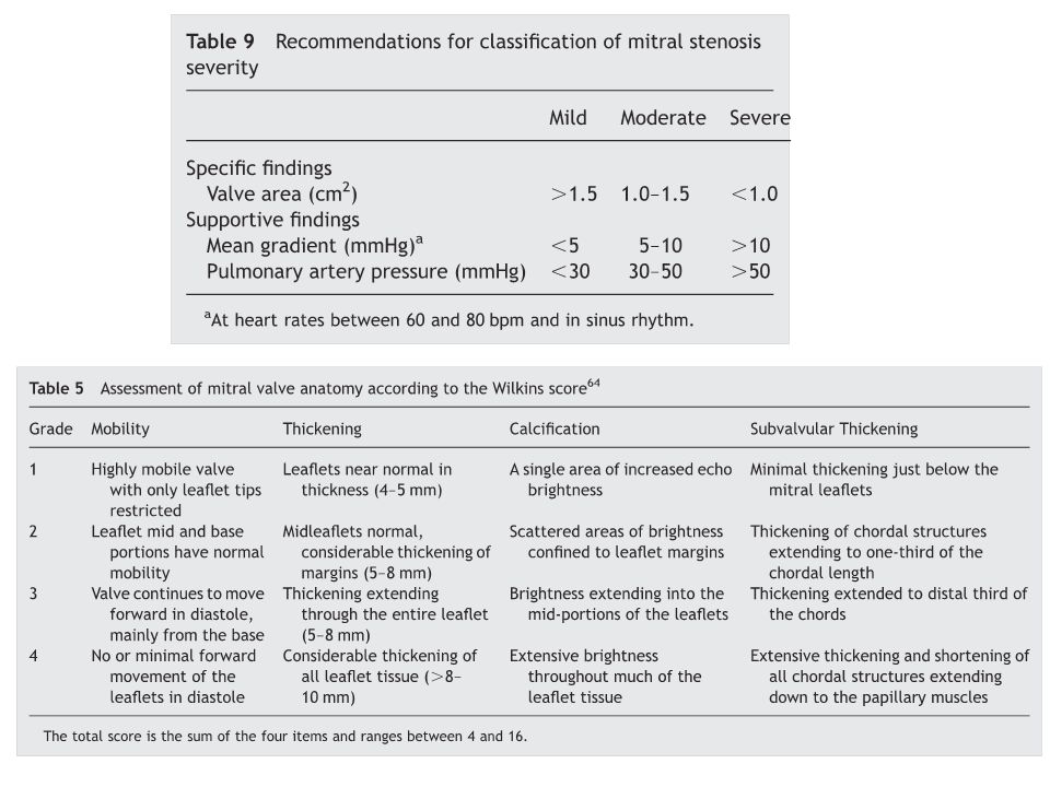
MS Ernstige MVA < 1.0cm2 en gradient >12mmHg MVA = 220/PHT
Bij prothese obstructie of mismatch bij mean gradient >10mmHg en PHT >160ms

Verwante presentaties


![Dick Ket [1902 – 1940]. Dick Ket [1902 – 1940]](/8/2134367/big_thumb.jpg "Dick Ket [1902 – 1940]. Dick Ket [1902 – 1940]>")


>")


