Download de presentatie
De presentatie wordt gedownload. Even geduld aub

1
TRANSPORT 6v

2
BS 1: Bloedsomloop Omloop Versimpelde versie Realistische versie

3
Bloedsomloop Aders Slagaders Enkele bloedsomloop Dubbele bloedsomloop
Meestal zuurstofarm Slagaders Meestal zuurstofrijk Enkele bloedsomloop 1 keer door het hart Dubbele bloedsomloop 2 keer door het hart Kleine bloedsomloop Grotebloedsomloop Bloedsomloop van een vis

4
Bij insecten

5
BS 2: Bloed Bloedplasma Bloedcellen Bloedplaatjes Functie algemeen
Rode bloedcellen Witte bloedcellen Bloedplaatjes Functie algemeen Vervoeren van stoffen Zuurstof (Rode bloedcellen) Voedingsstoffen (bloedplasma)
 Voedingsstoffen (bloedplasma)")
6
Bloedplasma Samenstelling Vervoer Fibrinogeen Plasma-eitwitten Water
Opgeloste stoffen (zouten) Vervoer Zuurstof (zeer weinig) Voedingsstoffen Koolstofdioxide Afvalstoffen (lever) Fibrinogeen Fibrine Eiwit Stolling Dichting van wonden
 Vervoer. Zuurstof (zeer weinig) Voedingsstoffen. Koolstofdioxide. Afvalstoffen (lever) Fibrinogeen. Fibrine. Eiwit. Stolling. Dichting van wonden.")
7
Bloedbestanddelen Withdraw blood and place in tube Centrifuge
Plasma (55% of whole blood) Buffy coat: leukocyctes and platelets (<1% of whole blood) Formed elements Erythrocytes (45% of whole blood) 1 Withdraw blood and place in tube 2 Centrifuge Figure 17.1
 Buffy coat: leukocyctes and platelets (<1% of whole blood) Formed elements. Erythrocytes (45% of whole blood) 1. Withdraw blood and place in tube. 2. Centrifuge. Figure")
8
Samenstelling van Bloed
Bloed is vloeibaar weefsel Het bestaat uit: plasma (vloeistof) Gevormde elementen: Erythrocytes, or red blood cells (RBCs) Leukocytes, or white blood cells (WBCs) Plaatjes Hematocriet = het percentage dat het volume rode bloedcellen in het bloed inneemt. Ongeveer 45 %
 Gevormde elementen: Erythrocytes, or red blood cells (RBCs) Leukocytes, or white blood cells (WBCs) Plaatjes. Hematocriet = het percentage dat het volume rode bloedcellen in het bloed inneemt. Ongeveer 45 %")
9
Fysiologische eigenschappen bloed
Bloed is plakkerig, ondoorzichtig en smaakt naar metaal De kleur varieert van licht rood tot donker rood De pH van bloed is 7.35–7.45 Temperatuur is 38C, ietsje hoger dan de lich.temp Het bloedvolume = 8% van het lichaamsgewicht Gemiddeld is 5–6 L voor de man, and 4–5 L voor de vrouw

10
Bloed Plasma Bloed plasma bevat 100 opgeloste en niet-opgeloste deeltjes: Eiwitten: albumine, globulines, stollingseiwitten enz Zouten Hormonen Organische verbindingen: voedingsstoffen, bouwstoffen enz. Gassen: zuurstof, koolstofdioxide, stikstof enz.

11
Overzicht bloedcirculatie 1
Bloed stroomt van hart via slagaders naar haarvaten Oxygen (O2) en voedingsstoffen diffunderen door de haarvaten wand naar de weefsels Carbon dioxide (CO2) en afval gaan van weefsel naar bloed
 en voedingsstoffen diffunderen door de haarvaten wand naar de weefsels. Carbon dioxide (CO2) en afval gaan van weefsel naar bloed.")
12
Overzicht bloedcirculatie 2
Zuurstof-arm bloed stroomt van haarvaten via aders naar hart In longen wordt CO2 afgestaan en O2 opgenomen Zuurstofrijk bloed gaat van longen terug naar LB

13
Taken van het bloed: transport
Bloed transports: Zuurstof van.. Naar.. Afvalproducten van de stofwisseling van.. naar Hormonen van klieren naar doelorganen

14
Taken bloed: Regulatie
Bloed zorgt voor: Handhaving lichaamstemperatuur Normale pH in weefsels, ondanks stofwisseling (buffer) Juiste vloeistofvolume in weefsels en bloedvaten
 Juiste vloeistofvolume in weefsels en bloedvaten.")
15
Taken bloed: verdediging
Bloed voorkomt bloedverlies door: Activering bloedplaatjes (stolling) Bloedpropvorming bij beschadiging bloedvat Bloed voorkomt infectie door: Productie en gebruik antistoffen door b-lymfocyten Activering complementsysteem in plasma Activering fagocytose door witte bloedcellen
 Bloedpropvorming bij beschadiging bloedvat. Bloed voorkomt infectie door: Productie en gebruik antistoffen door b-lymfocyten. Activering complementsysteem in plasma. Activering fagocytose door witte bloedcellen.")
16
Erythrocytes (RBCs) Biconcave schijfjes, kernloos!, belangrijke celorganellen ontbreken => sterven binnen 4 maanden! Bevatten hemoglobine, eiwit dat zuurstof kan binden en vervoeren.

17
Erythrocytes (RBCs) Figure 17.3
 Figure 17.3")
18
Structure of Hemoglobin
Figure 17.4

19
Vormen van Hemoglobine
Oxyhemoglobine – hemoglobine waaraan zuurstof is verbonden. Dit is GEEN OXIDATIE maar OXIGENATIE Zuurstof opname in de longen Desoxyhemoglobin – hemoglobin dat zuurstof in weefsels heeft afgegeven. (gereduceerde Hb) Carbaminohemoglobine – hemoglobine waaraan koolstofdioxide is verbonden Koolstofdioxide wordt opgenomen in de weefsels
 Carbaminohemoglobine – hemoglobine waaraan koolstofdioxide is verbonden. Koolstofdioxide wordt opgenomen in de weefsels.")
20
Figure 17.14

21
Bloedplaatjes (trombocytes)
Plaatjes zijn celfragmenten: uit elkaar gevallen megakaryocytes . Plaatjes funktie: propvorming voor tijdelijke afsluiting lekken bloedvat.

22
Vorming bloedplaatjes
De stamcel van het bloedplaatje is: de hemocytoblast Figure 17.12

23
Hemostasis = bloedstelping
Kettingreactie stopt het bloeden 3 fasen: Vaat contractie – samentrekking beschadigd bloedvat Bloedplaatjes vormen een draderig netwerk Coagulation (bloedcellen lopen vast in net) Filmpje
 Filmpje.")
24
Coagulation (Stolling)
Figure 17.13a

25
Stolling in 3 stappen Fase 1: Prothrombine Activator vorming
Fase 2: Prothrombin activator stimuleert omzetting prothrombin in actief enzym: thrombine Fase 3: Thrombine stimuleert de polymerizatie van fibrinogeen in fibrine Korstvorming en herstel van weefel Samentrekking fibrine draden => serum wordt eruit geperst Herstel: Onder korste worden o.i.v. weefselhormonen de huidgestimuleerd te delen en te herstellen Stolling-problemen Stollingsziekten Spontane stolling => trombose Hoe wordt dit voorkomen

26
BS 3: Het Hart Cardiac muscle bundles

27
Bouw van het hart, achteraanzicht.
Aorta Bovenste holle ader Linker long sl.a Rechter long sl.a. L longaders Rechter longader Oortje L boezem Rechter boezem Linker boezem Onderste holle ader kransader Rechter kranssl.a Kransader linker kamer Krans sinus Krans sl. a. anterieur Linker kamer Apex (d) Rechter kamer
 Rechter kamer.")
28
Overzicht hart, doorsnede
Aorta Bovenste holle ader Linker sla. Rechter long sl.a. Linker boezem Long sl.a. Linker long sl.a. Rechter boezem Rechter long aders Mitral (hart) klep (2 hoofdig) Aortic valve Halve maan vormig klep Longsl.a. Hartklep (3 hoofdig) Liner kamer Aanhechting spiertje Rechter kamer Pezen hartklep tussenschot hartspier trabekeltjes pericard Onderste Holle ader Endocardium = binnen Bekleding hart (e)
 klep. (2 hoofdig) Aortic. valve. Halve maan vormig klep. Longsl.a. Hartklep. (3 hoofdig) Liner kamer. Aanhechting spiertje. Rechter kamer. Pezen hartklep. tussenschot. hartspier. trabekeltjes. pericard. Onderste. Holle ader. Endocardium = binnen. Bekleding hart. (e)")
29
Doorsnede hart LK RK hartspier

30
Dubbele bloedsomloop. longen Kleine bloedsomloop Long sl.a. longader
Aorta Holle aders LB LK RB Heart RK Grote bloedsomloop Key: haarvatenbed = zuurstof rijk CO2 arm = zuurstof arm, CO2-rijk

31
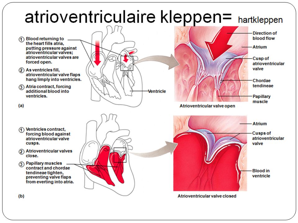
Hartkleppen, bovenaanzicht
Halve maanv.kleppen long.sl.a. Aortickleppejn Weggesnede deel Hartklep LK Hartklep RK hartspier Tricuspid) klep Mitraal klep Aorta klep longsla kleppen (b) Bindweefsel baan Anterior (a)
 klep. Mitraal. klep. Aorta. klep. longsla. kleppen. (b) Bindweefsel. baan. Anterior. (a)")
32
Hartkleppen Opening of superior vena cava Mitral valve Chordae
tendineae Tricuspid valve Myocardium of right ventricle Interventricular septum Papillary muscles Myocardium of left ventricle Chordae tendineae attached to tricuspid valve flap Papillary muscle (d) Pulmonary valve Aortic valve (c) Area of cutaway Mitral valve Tricuspid valve
 Pulmonary valve. Aortic valve. (c) Area of cutaway. Mitral valve. Tricuspid valve.")
34
Halvemaanvormige kleppen
As ventricles relax and intraventricular pressure falls, blood flows back from arteries, filling the cusps of semilunar valves and forcing them to close. As ventricles contract and intraventricular pressure rises, blood is pushed up against semilunar valves, forcing them open. Aorta Pulmonary trunk Semilunar valve open Semilunar valve closed (a) (b)
 (b)")
35
Pacemakercellen produceren zelfstandig (onafhankelijk van een zenuwprikkel!!) een AP(actiepotentiaal) (in sinusknoop) Ca2+ channels close; +10 K+ channels open –10 Ca2+ permeability –20 Action potential Ca2+ channels open K+ perme- ability –30 Membrane potential (mV) –40 –50 –60 Slow depolarization: Pacemaker potential –70 K+ channels close; slow Na+ channels opening (Na+ enters) Threshold Time (ms)
 –40. –50. –60. Slow depolarization: Pacemaker potential. –70. K+ channels close; slow Na+ channels. opening (Na+ enters) Threshold. Time (ms)")
36
Voortgeleiding AP die leidt tot contractie hartspieren
Superior vena cava Right atrium SA node 1 Sinoatrial (SA) node (pacemaker) Left atrium Internodal pathway Atrial muscle 2 Atrioventricular (AV) node 3 Atrioventricular (AV) bundle (Bundle of His) Purkinje fibers AV node Ventricular muscle 4 Bundle branches Inter- ventri- cular septum 5 Purkinje fibers 100 200 300 400 Milliseconds (a) (b)
 node (pacemaker) Left atrium. Internodal. pathway. Atrial muscle. 2. Atrioventricular. (AV) node. 3. Atrioventricular. (AV) bundle. (Bundle of His) Purkinje. fibers. AV node. Ventricular. muscle. 4. Bundle branches. Inter- ventri- cular. septum. 5. Purkinje fibers Milliseconds. (a) (b)")
37
Autonome hartslag regeling Nerver vagus => vertraging Nervus accellerans => versnelling
Dorsal motor nucleus of vagus Cardioinhibitory center (parasympathetic) Vagus nerve Cardioacceleratory center (sympathetic) Medulla oblongata Sympathetic trunk ganglion Thoracic spinal cord Sympathetic trunk Sympathetic cardiac nerve Key: Parasympathetic fibers Sympathetic fibers Interneurons AV node SA node
 Vagus. nerve. Cardioacceleratory. center (sympathetic) Medulla oblongata. Sympathetic. trunk. ganglion. Thoracic spinal cord. Sympathetic trunk. Sympathetic. cardiac. nerve. Key: Parasympathetic. fibers. Sympathetic. fibers. Interneurons. AV. node. SA. node.")
38
Electrocardiogram Animatie QRS complex Sinoatrial node R Ventricular
depolarization Atrioventricular node Ventricular repolarization Atrial depolarization T P Q P-Q Interval S-T Segment S Time (s) 0.2 0.4 0.6 0.8 Animatie Q-T Interval
 Animatie. Q-T. Interval.")
39
Relatie ECG contractie verloop
SA node generates impulse; atrial excitation begins Impulse delayed at AV node Impulse passes to heart apex; ventricular excitation begins Ventricular excitation complete AV node Bundle branches Purkinje fibers SA node

40
BS 4: Bloedvaten Transportbaan = bloedvatenstelsel
Transportmotor = hart Transportmiddel = bloed

41
Taken van het bloed onderhoud Homeostase TRANSPORT van Electrolyten
O2 & CO2 Afvalproducten Hormonen eiwitten Voedingsstoffen VERDEDIGING Vreemde organismen Verwonding infectie Stolling Lichaamstemperatuur onderhoud Homeostase

42
Bloedvat met bloed Witte bloedcel Rode bloedcel wand bloedplaatje

43
Typen bloedvaten slagader ader (a) Tunica intima klep • Endotheel •
Subendotheel laag Interne elastische lamina Tunica media= spierlaag External elastic lamina Tunica externa= bwschede Lumen Lumen Sl.a. Capillair network ader Endothelial cells (b) Capillair
 Capillair.")
44
Kenmerken slagader / ader
Slagader arterie Dikke gespierde wand Liggen diep Hoge bloeddruk Kloppen Geen kleppen Ader vene Dunne niet-gespierde wand Liggen aan het oppervlak Lage bloeddruk Kloppen niet Wel kleppen

45
Kenmerken van de slagader, ader en haarvaten

46
Temporal artery Facial artery Common carotid artery Brachial artery
Figure 19.11: Body sites where the pulse is most easily palpated, p. 732. Temporal artery Facial artery Common carotid artery Brachial artery Radial artery Femoral artery Popliteal artery Posterior tibial artery Dorsalis pedis artery

48
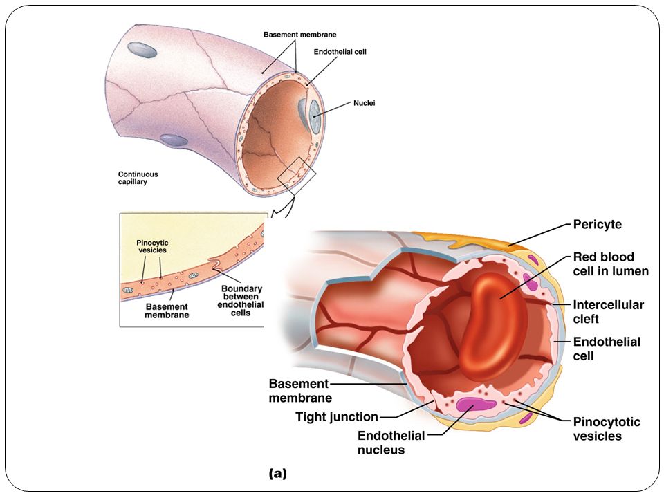
Uitwisseling haarvaten <> weefsel
Pericyt Lumen Pinocytotic vesicles porie Intercellulair ruimte Pinocytose blaasjes Rode bloed cel in lumen 4 Transport via blaasjes (grote mol.) Vensters= porien) Basement membrane Intercellular ruimte 2 Beweging door spleet (water- oplosbare stoffen) 3 Beweging door porie (water-soluble substances) Endotheel cel: kern 1 Diffusie door membraan (vetoplosbare stoffen) Basaal membraan Tight junction Endotheel cel
 Vensters= porien) Basement. membrane. Intercellular. ruimte. 2. Beweging door. spleet (water- oplosbare stoffen) 3. Beweging door. porie. (water-soluble. substances) Endotheel cel: kern. 1. Diffusie. door. membraan. (vetoplosbare. stoffen) Basaal membraan. Tight junction. Endotheel. cel.")
49
Pinocytose blaasjes Pericyt Intercellulaire ruimte Rode bloedcel
Bouw capillair Pericyt Pinocytose blaasjes Intercellulaire ruimte Rode bloedcel Endotheel cel Venster/porie Basaal membrane Endotheel kern Intercellulaire ruimte Tight junction Pinocytose blaasjes Endotheel kern (a) (b) (c)
 (b) (c)")
50
Bloed verdeling Veneus systeem Arterieel systeem Grote aders Hart
Elastische arterien grote lymfe vaten Capacitance vessels lymfe knoop Gespierde arterien (distributing vessels) Lymphatisch system Kleine aders Arterioveneuze anastomose Lymphatische capillairen Arteriolen (weerstand!) Sinus= verwijding Precapillaire sphincter shunt (a) Haarvaten gaswisseling Bloedvaten longen 12% Hart 8% Ateriele bloedvaten 15% Veneuze bloed- vaten 60% (b) Capillarien 5%
 Lymphatisch. system. Kleine aders. Arterioveneuze. anastomose. Lymphatische. capillairen. Arteriolen. (weerstand!) Sinus= verwijding. Precapillaire. sphincter. shunt. (a) Haarvaten. gaswisseling. Bloedvaten longen 12% Hart 8% Ateriele bloedvaten 15% Veneuze bloed- vaten. 60% (b) Capillarien 5%")
51
Bloedverdeling (Diagram)
")
52
slagader-haarvat-ader doorbloeding geregeld via precapilaire kringspiertjes
Vasculaire shunt Precapillaire sphincters hoofdcapillair (a) Sphincter open (b) Sphincters gesloten
 Sphincter open. (b) Sphincters gesloten.")
53
BS 5: Bloeddruk Bloeddruk bepaald door: Perifere weerstand Bloedvolume
Slagkracht hart Slagvolume hart

54
Regeling bloeddruk Figure 19.8 Impulse traveling along
afferent nerves from baroreceptors: Stimulate cardio- inhibitory center (and inhibit cardio- acceleratory center) Sympathetic impulses to heart ( HR and contractility) Baroreceptors in carotid sinuses and aortic arch stimulated Inhibit vasomotor center CO R Rate of vasomotor impulses allows vasodilation ( vessel diameter) Arterial blood pressure rises above normal range CO and R return blood pressure to Homeostatic range Stimulus: Rising blood pressure Imbalance Homeostasis: Blood pressure in normal range Stimulus: Declining blood pressure Imbalance CO and R return blood pressure to homeostatic range Impulses from baroreceptors: Stimulate cardio- acceleratory center (and inhibit cardio- inhibitory center) Arterial blood pressure falls below normal range Cardiac output (CO) Baroreceptors in carotid sinuses and aortic arch inhibited Sympathetic impulses to heart ( HR and contractility) Peripheral resistance (R) Vasomotor fibers stimulate vasoconstriction Stimulate vasomotor center Figure 19.8
 Sympathetic. impulses to. heart. ( HR and contractility) Baroreceptors. in carotid. sinuses and. aortic arch. stimulated. Inhibit. vasomotor center. CO. R. Rate of vasomotor. impulses allows. vasodilation. ( vessel diameter) Arterial. blood pressure. rises above. normal range. CO and R. return blood. pressure to. Homeostatic. range. Stimulus: Rising blood. pressure. Imbalance. Homeostasis: Blood pressure in normal range. Stimulus: Declining. blood pressure. Imbalance. CO and R. return blood. pressure to. homeostatic. range. Impulses from. baroreceptors: Stimulate cardio- acceleratory center. (and inhibit cardio- inhibitory center) Arterial blood pressure. falls below normal range. Cardiac. output. (CO) Baroreceptors in. carotid sinuses. and aortic arch. inhibited. Sympathetic. impulses to heart. ( HR and contractility) Peripheral. resistance (R) Vasomotor. fibers. stimulate. vasoconstriction. Stimulate. vasomotor. center. Figure")
55
Homeostasis: Blood pressure in normal range
Figure 19.8

56
Figure 19.8 Stimulus: Rising blood Imbalance pressure
Homeostasis: Blood pressure in normal range Imbalance Figure 19.8

57
Figure 19.8 Baroreceptors in carotid sinuses and aortic arch
stimulated Arterial blood pressure rises above normal range Stimulus: Rising blood pressure Imbalance Homeostasis: Blood pressure in normal range Imbalance Figure 19.8

58
Figure 19.8 Impulse traveling along afferent nerves from
baroreceptors: Stimulate cardio- inhibitory center (and inhibit cardio- acceleratory center) Baroreceptors in carotid sinuses and aortic arch stimulated Inhibit vasomotor center Arterial blood pressure rises above normal range Stimulus: Rising blood pressure Imbalance Homeostasis: Blood pressure in normal range Imbalance Figure 19.8
 Baroreceptors. in carotid. sinuses and. aortic arch. stimulated. Inhibit. vasomotor center. Arterial. blood pressure. rises above. normal range. Stimulus: Rising blood. pressure. Imbalance. Homeostasis: Blood pressure in normal range. Imbalance. Figure")
59
Figure 19.8 Impulse traveling along afferent nerves from
baroreceptors: Stimulate cardio- inhibitory center (and inhibit cardio- acceleratory center) Sympathetic impulses to heart ( HR and contractility) Baroreceptors in carotid sinuses and aortic arch stimulated Inhibit vasomotor center Rate of vasomotor impulses allows vasodilation ( vessel diameter) Arterial blood pressure rises above normal range Stimulus: Rising blood pressure Imbalance Homeostasis: Blood pressure in normal range Imbalance Figure 19.8
 Sympathetic. impulses to. heart. ( HR and contractility) Baroreceptors. in carotid. sinuses and. aortic arch. stimulated. Inhibit. vasomotor center. Rate of vasomotor. impulses allows. vasodilation. ( vessel diameter) Arterial. blood pressure. rises above. normal range. Stimulus: Rising blood. pressure. Imbalance. Homeostasis: Blood pressure in normal range. Imbalance. Figure")
60
CO = cardiac output = hartslag volume R = weerstand
HR = hartslagfrequentie Impulse traveling along afferent nerves from baroreceptors: Stimulate cardio- inhibitory center (and inhibit cardio- acceleratory center) Sympathetic impulses to heart ( HR and contractility) Baroreceptors in carotid sinuses and aortic arch stimulated Inhibit vasomotor center CO R Rate of vasomotor impulses allows vasodilation ( vessel diameter) Arterial blood pressure rises above normal range CO and R return blood pressure to homeostatic range Stimulus: Rising blood pressure Homeostasis: Blood pressure in normal range Figure 19.8
 Sympathetic. impulses to. heart. ( HR and contractility) Baroreceptors. in carotid. sinuses and. aortic arch. stimulated. Inhibit. vasomotor center. CO. R. Rate of vasomotor. impulses allows. vasodilation. ( vessel diameter) Arterial. blood pressure. rises above. normal range. CO and R. return blood. pressure to. homeostatic. range. Stimulus: Rising blood. pressure. Homeostasis: Blood pressure in normal range. Figure")
61
Figure 19.8 Imbalance Homeostasis: Blood pressure in normal range
Stimulus: Declining blood pressure Imbalance Figure 19.8

62
Figure 19.8 Imbalance Homeostasis: Blood pressure in normal range
Stimulus: Declining blood pressure Imbalance Impulses from baroreceptors: Stimulate cardio- acceleratory center (and inhibit cardio- inhibitory center) Arterial blood pressure falls below normal range Baroreceptors in carotid sinuses and aortic arch inhibited Figure 19.8
 Arterial blood pressure. falls below normal range. Baroreceptors in. carotid sinuses. and aortic arch. inhibited. Figure")
63
Figure 19.8 Imbalance Homeostasis: Blood pressure in normal range
Stimulus: Declining blood pressure Imbalance Impulses from baroreceptors: Stimulate cardio- acceleratory center (and inhibit cardio- inhibitory center) Arterial blood pressure falls below normal range Baroreceptors in carotid sinuses and aortic arch inhibited Stimulate vasomotor center Figure 19.8
 Arterial blood pressure. falls below normal range. Baroreceptors in. carotid sinuses. and aortic arch. inhibited. Stimulate. vasomotor. center. Figure")
64
Figure 19.8 Imbalance Homeostasis: Blood pressure in normal range
Stimulus: Declining blood pressure Imbalance Impulses from baroreceptors: Stimulate cardio- acceleratory center (and inhibit cardio- inhibitory center) Arterial blood pressure falls below normal range Baroreceptors in carotid sinuses and aortic arch inhibited Sympathetic impulses to heart ( HR and contractility) Vasomotor Fibers stimulate vasoconstriction Stimulate vasomotor center Figure 19.8
 Arterial blood pressure. falls below normal range. Baroreceptors in. carotid sinuses. and aortic arch. inhibited. Sympathetic. impulses to heart. ( HR and contractility) Vasomotor. Fibers. stimulate. vasoconstriction. Stimulate. vasomotor. center. Figure")
65
Figure 19.8 Homeostasis: Blood pressure in normal range Stimulus:
Declining blood pressure CO and R return blood pressure to homeostatic range Impulses from baroreceptors: Stimulate cardio- acceleratory center (and inhibit cardio- inhibitory center) Arterial blood pressure falls below normal range Cardiac output (CO) Baroreceptors in carotid sinuses and aortic arch inhibited Sympathetic impulses to heart ( HR and contractility) Peripheral resistance (R) Vasomotor Fibers stimulate vasoconstriction Stimulate vasomotor center Figure 19.8
 Arterial blood pressure. falls below normal range. Cardiac. output. (CO) Baroreceptors in. carotid sinuses. and aortic arch. inhibited. Sympathetic. impulses to heart. ( HR and contractility) Peripheral. resistance (R) Vasomotor. Fibers. stimulate. vasoconstriction. Stimulate. vasomotor. center. Figure")
66
DUS: als bloeddruk te hoog wordt…
CO = cardiac output = hartslag volume R = weerstand HR = hartslagfrequentie Impulse traveling along afferent nerves from baroreceptors: Stimulate cardio- inhibitory center (and inhibit cardio- acceleratory center) Sympathetic impulses to heart ( HR and contractility) Baroreceptors in carotid sinuses and aortic arch stimulated Inhibit vasomotor center CO= cardiac output = slagvolume R Arterial blood pressure rises above normal range Rate of vasomotor impulses allows vasodilation ( vessel diameter) CO and R return blood pressure to homeostatic range Stimulus: Rising blood pressure Imbalance Homeostasis: Blood pressure in normal range Imbalance
 Sympathetic. impulses to. heart. ( HR and contractility) Baroreceptors. in carotid. sinuses and. aortic arch. stimulated. Inhibit. vasomotor center. CO= cardiac output = slagvolume. R. Arterial. blood pressure. rises above. normal range. Rate of vasomotor. impulses allows. vasodilation. ( vessel diameter) CO and R. return blood. pressure to. homeostatic. range. Stimulus: Rising blood. pressure. Imbalance. Homeostasis: Blood pressure in normal range. Imbalance.")
67
Dus als bloeddruk te laag wordt…..
CO = cardiac output = hartslag volume R = weerstand HR = hartslagfrequentie Imbalance Homeostasis: Blood pressure in normal range Stimulus: Declining blood pressure Imbalance CO and R return blood pressure to Homeostatic range Impulses from baroreceptors: Stimulate cardio- acceleratory center (and inhibit cardio- inhibitory center) Arterial blood pressure falls below normal range Cardiac output (CO) Baroreceptors in carotid sinuses and aortic arch inhibited Sympathetic impulses to heart ( HR and contractility) Peripheral resistance (R) Vasomotor fibers stimulate vasoconstriction Stimulate vasomotor center
 Arterial blood pressure. falls below normal range. Cardiac. output. (CO) Baroreceptors in. carotid sinuses. and aortic arch. inhibited. Sympathetic. impulses to heart. ( HR and contractility) Peripheral. resistance (R) Vasomotor. fibers. stimulate. vasoconstriction. Stimulate. vasomotor. center.")
68
Figure 19.8 Impulse traveling along afferent nerves from
baroreceptors: Stimulate cardio- inhibitory center (and inhibit cardio- acceleratory center) Sympathetic impulses to heart ( HR and contractility) Baroreceptors in carotid sinuses and aortic arch stimulated Inhibit vasomotor center CO R Rate of vasomotor impulses allows vasodilation ( vessel diameter) Arterial blood pressure rises above normal range CO and R return blood pressure to Homeostatic range Stimulus: Rising blood pressure Imbalance Homeostasis: Blood pressure in normal range Stimulus: Declining blood pressure Imbalance CO and R return blood pressure to homeostatic range Impulses from baroreceptors: Stimulate cardio- acceleratory center (and inhibit cardio- inhibitory center) Arterial blood pressure falls below normal range Cardiac output (CO) Baroreceptors in carotid sinuses and aortic arch inhibited Sympathetic impulses to heart ( HR and contractility) Peripheral resistance (R) Vasomotor fibers stimulate vasoconstriction Stimulate vasomotor center Figure 19.8
 Sympathetic. impulses to. heart. ( HR and contractility) Baroreceptors. in carotid. sinuses and. aortic arch. stimulated. Inhibit. vasomotor center. CO. R. Rate of vasomotor. impulses allows. vasodilation. ( vessel diameter) Arterial. blood pressure. rises above. normal range. CO and R. return blood. pressure to. Homeostatic. range. Stimulus: Rising blood. pressure. Imbalance. Homeostasis: Blood pressure in normal range. Stimulus: Declining. blood pressure. Imbalance. CO and R. return blood. pressure to. homeostatic. range. Impulses from. baroreceptors: Stimulate cardio- acceleratory center. (and inhibit cardio- inhibitory center) Arterial blood pressure. falls below normal range. Cardiac. output. (CO) Baroreceptors in. carotid sinuses. and aortic arch. inhibited. Sympathetic. impulses to heart. ( HR and contractility) Peripheral. resistance (R) Vasomotor. fibers. stimulate. vasoconstriction. Stimulate. vasomotor. center. Figure")
Verwante presentaties


 –Dubbele bloedsomloop Grote.>")


