Download de presentatie
De presentatie wordt gedownload. Even geduld aub

1
16 – 04 – 2014 Tessa Biesheuvel AFAS stadion Congres traumatologie
Thorax en abdomen 16 – 04 – 2014 Tessa Biesheuvel AFAS stadion Congres traumatologie

3
Thoraxletsel ?

5
Thorax Trauma Significante oorzaak mortaliteit
< 10% operatief ingrijpen Verschil in scherp en stomp letsel Bij scherp letsel 15-30% operatie Meeste patiënten simpele procedures 4-5 Thoracic Trauma Review this information with the students. Emphasize that chest injuries remain a significant cause of morbidity and mortality among trauma patients. Life-threatening injuries associated with thoracic injuries are identified in the primary survey by carefully assessing the patient’s ABCs. Identified injuries usually require simple interventions to secure the airway, reexpand the lung, drain the pleural space, and improve breathing mechanics. Most penetrating wounds to the chest require a thoracostomy tube. A minority of patients with chest injuries require urgent surgical exploration due to bleeding. Use the final point to elicit the six life threatening injuries on the subsequent slide.

7
Waar op te letten ? Look Listen Feel

8
Waar op te letten ? ABCD LOOK: Hematomen / wonden ?
Thoraxwand deformatie ? Ademhalingsfrequentie Hulpademhalingsspieren / “respiratoire nood” ? Neusvleugelen Trachea mediaan Nekvenen Sputum

9
Vervolg, waar op letten ? LISTEN: Stridor Andere percussie
Verminderd ademgeruis FEEL: Symmetrische ademexcusies Subcutaan emfyseem Crepitaties Pijn

10
5 levensbedreigende thoraxletsel
The big 5 5 levensbedreigende thoraxletsel

11
The big 5 A B Spanningspneumothorax Open pneumothorax
Fladder thorax en Long contusie C Massieve hematothorax Cardiale tamponade

12
Spanningspneumothorax
Onrust Shock Uitgezette nekvenen AG Hypersonoor Cyanose (laat !) 4-9 Tension Pneumothorax How do I identify a tension pneumothorax? How do I differentiate a tension pneumothorax from cardiac tamponade and hemorrhagic shock? During the discussion of differentiating a tension pneumothorax from cardiac tamponade and hemorrhagic shock, relate that blunt injury is a more likely cause of tension pneumothorax. Cardiac tamponade occurs more often with penetrating trauma. If hypovolemia exists, the patient’s neck veins will not be distended.
 4-9 Tension Pneumothorax. How do I identify a tension pneumothorax How do I differentiate a tension pneumothorax from cardiac tamponade and hemorrhagic shock During the discussion of differentiating a tension pneumothorax from cardiac tamponade and hemorrhagic shock, relate that blunt injury is a more likely cause of tension pneumothorax. Cardiac tamponade occurs more often with penetrating trauma. If hypovolemia exists, the patient’s neck veins will not be distended.")
13
Fladderthorax en Longcontusie
Zuurstof Reëxpanderen long Intuberen Matig infuus Pijnstilling 4-14 Flail Chest and Pulmonary Contusion How do I treat the patient with a flail chest and/or pulmonary contusion? Emphasize that the treatment goal is to reexpand the lung, eg, with CPAP (positive pressure) or physiotherapy, and to avoid progressive atelectasis. Analgesia is an important adjunct, but oversedation will promote hypoventilation and atelectasis.
 or physiotherapy, and to avoid progressive atelectasis. Analgesia is an important adjunct, but oversedation will promote hypoventilation and atelectasis.")
14
Massieve Hematothorax
Systemische - / long vaten > 1500 mL bloed- verlies Platte nekvenen Shock zonder AG en met gedempte percussie 4-15 Massive Hemothorax What is the cause and how do I identify if the patient has a massive hemothorax? Note that this type of injury results in a ‘B’ and ‘C’ problem. X-ray courtesy of Ray McGlone, Royal Lancaster Infirmary; UK

15
Hart Tamponade pols Uitgezette nekvenen Zachte harttonen PEA
4-17 Cardiac Tamponade This photograph shows a patient with a parasternal penetrating injury, self-inflicted with a car radio antenna. (Note: The patient’s head is at the bottom of the photograph where one can see the oxygen mask.) Ensure that the students understand that blunt trauma to the chest can cause cardiac tamponade, but that most survivors of cardiac tamponade have an anterior or posterior penetrating wound to the chest. During the discussion about signs and symptoms of the patient with cardiac tamponade, emphasize the fact that not all findings must be present. Explain that cardiac tamponade is suspected if the patient has a penetrating parasternal wound. Clinical signs may include hypotension and dyspnea, or the patient verbalizing that he or she senses he or she is dying. Photograph used with permission of Trauma.org; Frederick Foss; chest0016a;
 Ensure that the students understand that blunt trauma to the chest can cause cardiac tamponade, but that most survivors of cardiac tamponade have an anterior or posterior penetrating wound to the chest. During the discussion about signs and symptoms of the patient with cardiac tamponade, emphasize the fact that not all findings must be present. Explain that cardiac tamponade is suspected if the patient has a penetrating parasternal wound. Clinical signs may include hypotension and dyspnea, or the patient verbalizing that he or she senses he or she is dying. Photograph used with permission of Trauma.org; Frederick Foss; chest0016a;")
16
Andere potentieel bedreigende letsels
Letsel aan de bronchiaal boom Simpele pneumothorax Longcontusie Hematothorax Corcontusie Aortaruptuur Oesophagusletsel Traumatisch diafragma letsel

17
Andere potentieel bedreigende letsels

18
Simpele Pneumothorax Stomp / Scherp Hypersonore percussie ↓ AG
Thoraxdrain 4-26 Simple Pneumothorax How do I identify and treat a simple pneumothorax? The students should understand that a pneumothorax can occur after blunt and penetrating injury to the chest, although it may not be apparent on physical examination. After describing how to identify and treat this injury, ask about the usefulness of obtaining a chest x-ray, which can help identify this injury. Remind students of the need to assess for subcutaneous emphysema. The presence of decreased breath sounds is not always a helpful indicator for a pneumothorax, especially if the patient is hyperventilating from pain or is in shock. The students should know that this injury is treated with tube thoracostomy.

19
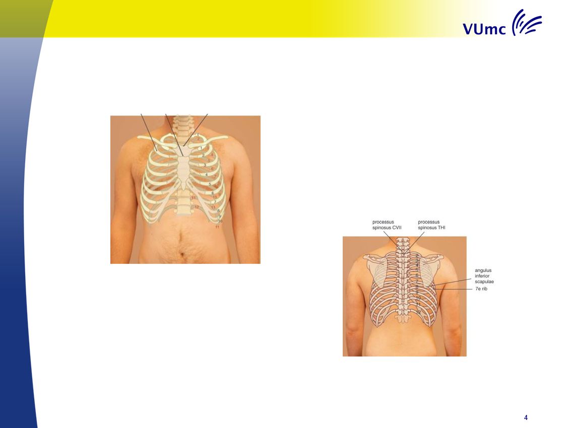
Fracturen rondom thorax en geassocieerd letsel
Sternum, Scapula, Rib Rib 1-3 Hoog energetisch Mortaliteit door onderliggend letsel Rib 4-9 Longcontusie en pneumothorax Rib 10-12 Denk aan buikletsel 4-37 Fractures and Associated Injuries What associated injuries should I suspect and assess for when my patient has fractures of the chest wall? Explain that the location of the fracture provides clues about other possible injuries. The students should understand that it requires a significant force to fracture ribs 1-3. Patients with these fractures commonly have many associated injuries and are at a higher risk for mortality. Fractures of ribs 4-9 are commonly associated with flail chest, pulmonary contusion, and pneumothorax. Lower rib fractures are associated with intraabdominal injury. Time permitting, review diaphragmatic excursion and its relationship to intraabdominal injuries and lower thoracic injuries. KINDEREN VRIJWEL NOOIT RIBFRACTUREN

20
Longcontusie VAAK ! Pijn, dyspneu, hemoptoë, hypoxie Oxygeneren
Eventueel beademen Late verandering op X thorax 4-27 Pulmonary Contusion How do I identify and treat a pulmonary contusion? A pulmonary contusion can be mild to severe and may cause very little hypoxia to severe hypoxia. The diagnosis can be confirmed by a chest x-ray or a CT scan of the chest. Most pulmonary contusions increase in size and severity after fluid resuscitation. The students should know that treatment includes normovolemia and maneuvers to maintain lung volumes. X-ray courtesy of Ray McGlone, Royal Lancaster Infirmary, UK

21
Samenvatting Niet zeldzaam Goed Kijken, Luisteren en voelen
Met simpele technieken grote pathologie te herkennen Denk aan onderliggende long vooral bij kinderen

Verwante presentaties
 after an unusually hard day on the job. You're really tired, and frustrated…… Vertaling:>")


gewone complicatie van een SAB>")