Download de presentatie
De presentatie wordt gedownload. Even geduld aub

1
Dag 3 Ischemie en infarct
Martijn Meuwissen Academisch Medisch Centrum, Amsterdam

2
non-profit / open access / physician moderated / up-to-date

3
Cursusoverzicht Dag 1: Basis, systematische beoordeling
Ivo van der Bilt Dag 2: Ritmestoornissen Jonas de Jong Dag 3: Ischemie Martijn Meuwissen De cursus is interactief. Onderbreek gerust!

4
Cardionetworks Auteurs: Jonas de Jong Ivo van der Bilt
Martijn Meuwissen Renée van den Brink Joris de Groot Illustraties: Rob Kreuger Bart Duineveld Met dank aan: Arthur Wilde Rudolph Koster Boeken: Wellens: The ECG in Emergency Decision Making Garcia / Miller: Arrhythmia Recognition Braunwald Heart Disease

5
Agenda Ischemie en Infarct
Achtergrond Diagnostiek Infarct localisatie Complicaties Quiz

6
Kort overzicht vorige week

7
Pathofysiologie Oorzaak acuut coronair syndroom = acute afname of blokkade in coronaire doorbloeding Atherosclerotische plaque Thrombotische occlusie tgv plaque ruptuur Plaatjes aggregatie Vasoactiva > spasme Thrombine > stollingsactivatie > bevorderen aggregatie Zelden: erosie/ embolie/ spasme/ anemie/ hypoxie

8
Pathofysiologie lumen lumen disrupted fibrous cap thin fibrous cap
Vulnerable Plaque lumen thin fibrous cap Stable Plaque thick fibrous cap disrupted fibrous cap Ruptured Plaque lumen adventitia intima thrombus media lipid core macrophages

9
Levensloop coronaire plaque
adhesie en migratie van ontstekingscellen 0% migratie van monocyten en gladde spier cellen 20% schuimcel vorming 30% atheroom en fibreuze cap vorming 50% “vulnerable plaque” – fragiele lesie 70% gecompliceerde lesie 90% tijd

10
Pathofysiologie Acuut myocard infarct
Enzymen (troponines, CK-MB) + 1 of meer van de volgende Ischaemische symptomen ECG afwijkingen passend bij ischaemie of necrose: ontwikkeling van Q-golven, ST Pathologische kenmerken van AMI (echo)
 + 1 of meer van de volgende. Ischaemische symptomen. ECG afwijkingen passend bij ischaemie of necrose: ontwikkeling van Q-golven, ST Pathologische kenmerken van AMI (echo)")
11
Grootte van infarct Demand vs supply: Supply: Demand
plaats afsluiting (proximaal/ distaal) duur afsluiting irreversibel na min collaterale doorbloeding (Hb, PO2) Demand zuurstof behoefte myocard frequentie, RR, contractiliteit
 duur afsluiting irreversibel na min. collaterale doorbloeding. (Hb, PO2) Demand. zuurstof behoefte myocard. frequentie, RR, contractiliteit.")
12
Collateraal vorming

13
35-day mortality reduction vs treatment delay
Waarom snelle infarctdiagnostiek? 35-day mortality reduction vs treatment delay 80 60 40 20 Absolute benefit per 1000 treated patients 3 6 9 12 15 18 21 24 Treatment delay (h) Boersma Lancet. 1996
 Boersma Lancet")
14
Patency of infarct-related comparing PTCA and Thrombolysis
CADILLAC GUSTO 1 TIMI 14 SP King III, Interventional Cardiology 2007

15
PRAGUE - 2 With the mortality on the y-axe, you can see that the overall mortality shows a difference in favor of PCI. Patients presented within 3 hours after symptom onset showed no difference in mortality between thrombolysis and PCI, the benefit is in patients presented more than 3 hours after onset of symptoms: 15.3% vs 6% This shows that long distance to a PCI center is safe and there is a decrease of mortality when presented more than 3 hours after onset of symptoms. STUDIE VOORTIJDIG GESTOPT VANWEGE BALK 3

16
Effect of door-to-door time on mortality
1.8 Relative risk of death Relative Risk of In-Hospital Death 1.6 1.4 1.2 1.0 McNamara et al. JACC 2006;47:

17
AHC / AHA Guidelines Primary PCI is “preferred treatment” for
ST-elevation acute myocardial infarction if: Symptoms < 12 hrs Time from “first medical contact” to start reperfusion therapy 90 min. Experienced team, high volume center

18
ESC Guidelines STEMI 2003 STEMI <12 hours after onset of chest pain
patient presenting in a hospital with PCI patient presenting in a hospital without PCI immediate transfer Thrombolysis ≥ 3 – 12 hours < 3 hours * failed successful PCI within up to 24 hours available PCI within up to 24 hours not available predischarge ischaemia primary PCI rescue PCI post thrombolysis PCI ischaemia-driven PCI

19
Decline in in-hospital mortality
S. Rasoul, Thesis 2007

20
Algemene Infarct Diagnostiek
Anamnese Drukkende retrosternale POB, dyspnoe, radiatie, angst, vegetatief, (geen) reactie op NTG, collaps , risicofactoren Lichamelijk onderzoek Vegetatieve verschijnselen, pols, bloeddruk, decompensatio cordis, shock ECG ST deviatie, Q’s, ritme-/ geleidingsstoornissen (LBTB) Lab troponine, CKMB etc
 reactie op NTG, collaps , risicofactoren. Lichamelijk onderzoek. Vegetatieve verschijnselen, pols, bloeddruk, decompensatio cordis, shock. ECG. ST deviatie, Q’s, ritme-/ geleidingsstoornissen (LBTB) Lab. troponine, CKMB etc.")
21
Algemene Diagnostiek Mogelijke gevolgen van AMI
Dyspneu: backward failure (MI?) Shock: forward failure (ruptuur?) Duizelig/collaps: ritme-/ geleidingsstoornissen (VF familieanamnese!) CVA: LV thrombus/ AFib NB: frequent geen (duidelijke) klachten ouderen, diabetici!!
 Shock: forward failure (ruptuur ) Duizelig/collaps: ritme-/ geleidingsstoornissen (VF familieanamnese!) CVA: LV thrombus/ AFib. NB: frequent geen (duidelijke) klachten ouderen, diabetici!!")
22
Differentiaal diagnostiek
Cardiaal (pericarditis, mechanische complicatie (stil) AMI, takotsubo) Vasculair Type A (B) dissectie, aneurysma Pulmonaal longembolie Gastro-oesophageaal spasme Bewegingsapparaat Tietze
 AMI, takotsubo) Vasculair. Type A (B) dissectie, aneurysma. Pulmonaal. longembolie. Gastro-oesophageaal. spasme. Bewegingsapparaat. Tietze.")
23
ECG Diagnostiek Acute coronair occlusie Instabiele angina pectoris
ST elevatie, (reciproke) depressie Q-vorming T-top inversie Geleiding: Nieuw linker- / rechter bundeltak blok As-draai QT verlenging Ritmestoornissen (VF/ AIVR/ AF/ totaal AVB) Instabiele angina pectoris Persisterende of voorbijgaande ST depressie T golf veranderingen (vlak / inversie / pseudo-normalisatie) Niet-specifiek / normaal ECG
 depressie. Q-vorming. T-top inversie. Geleiding: Nieuw linker- / rechter bundeltak blok. As-draai. QT verlenging. Ritmestoornissen (VF/ AIVR/ AF/ totaal AVB) Instabiele angina pectoris. Persisterende of voorbijgaande ST depressie. T golf veranderingen (vlak / inversie / pseudo-normalisatie) Niet-specifiek / normaal ECG.")
24
ECG infarct

25
ECG Diagnostiek Precordiaal

26
ECG Diagnostiek Precordiaal

27
ECG Diagnostiek

28
ECG Diagnostiek Extremiteiten

29
Systematische beoordeling
Algemene kenmerken Ritme Frequentie Geleidingstijden Hartas P top morfologie QRS morfologie ST morfologie Vergelijking met oud ECG Conclusie

30
7+2 STAPPENPLAN Stap 6: QRS morfologie
Pathologische Q top? Breedte ≥ 0.04 sec Diepte > ⅓ van de R Differentiaal diagnose? Oud infarct Cardiomyopathie (HCM, DCM) COPD Intraventriculaire geleidingsstoornissen
 COPD. Intraventriculaire geleidingsstoornissen.")
31
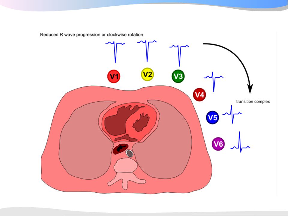
7+2 STAPPENPLAN Stap 6: QRS morfologie
R-top progressie? Overgangs complex in V3, V4 Normaal zit het overgangs complex (waar de R-top groter wordt dan de S) bij V3 tot V4
 bij V3 tot V4.")
32
7+2 STAPPENPLAN Stap 6: QRS morfologie
R-top progressie? Differentiaal diagnose onvoldoende r-top progressie? RV hypertrofie COPD, asthma Voorwand infarct of anteroseptaal infarct Geleidingsstoornissen (LBBB, Left anticus hemiblok, intraventriculaire geleidings vertraging) Cardiomyopathie Thorax afwijking Normale variant Precordiale afleidingen verkeerd geplaatst ANAMNESE EN LO/ ZIJN EXTREEM BELANGRIJK VOOR JUISTE INTERPRETATIE VAN HET ECG
 Cardiomyopathie. Thorax afwijking. Normale variant. Precordiale afleidingen verkeerd geplaatst. ANAMNESE EN LO/ ZIJN EXTREEM BELANGRIJK. VOOR JUISTE INTERPRETATIE VAN HET ECG.")
34
Vorm ST Segment concaaf of convex? B A

36
7+2 STAPPENPLAN Stap 7: ST-segment
Wanneer spreekt men van een pathologische ST elevatie? ST elevatie die op het J punt ≥ 1 mm is in afleiding I, II, III, aVL, aVF, V4-V6 ST elevatie die op het J punt ≥ 2 mm in V1-V3 ST elevatie treedt op bij: Transmurale ischemie of transmuraal infarct (STEMI) Differentiaal diagnose Pericarditis, LVH, brugada, takotsubo, K+, digitalis, bundeltakblok, CVA, hyperventilatie etc.
 Differentiaal diagnose. Pericarditis, LVH, brugada, takotsubo, K+, digitalis, bundeltakblok, CVA, hyperventilatie etc.")
37
Verschillende vormen van ST elevatie bij ischemie

38
Vroege Repolarisatie Zeer frequente bevinding “Smiley”configuratie
Overigens gezonde asymptomatische jonge volwassene Vaak in voorwands afl. Notching J punt Geen Q Geen reciproke ST depressie

39
Normale Varianten Figure 1. Electrocardiograms Showing Normal ST-Segment Elevation and Normal Variants. Tracing 1 shows normal ST-segment elevation. Approximately 90 percent of healthy young men have ST-segment elevation of 1 to 3 mm in one or more precordial leads. The ST segment is concave. Tracing 2 shows the early-repolarization pattern, with a notch at the J point in V4. The ST segment is concave, and the T waves are relatively tall. Tracing 3 shows a normal variant that is characterized by terminal T-wave inversion. The QT interval tends to be short, and the ST segment is coved.

40
ST Varianten

41
7+2 STAPPENPLAN Stap 7+1: Vergelijk met oud ECG
Verandering ritme? Nieuw boezemfibrilleren? Verandering frequentie? Bradycardie (SB, AV blokmedicatie effect?) of tachycardie ([S]VT) Verandering geleidingstijden? PQ tijd (medicatie?); QRS (ischemie, medicatie?) QT tijd (medicatie?) Verandering hartas? Verandering geleiding, infarct doorgemaakt? Nieuwe pathologische Q’s Verandering geleiding, infarct doorgemaakt, plaatsing elektroden? Verandering R top progressie precordiaal? Afname R (infarct, tamponade, plaatsing elektroden?) Toename R top (LVH, RVH, verandering geleiding intraventriculair) Verandering ST segment? Verandering T-top?
 of tachycardie ([S]VT) Verandering geleidingstijden PQ tijd (medicatie ); QRS (ischemie, medicatie ) QT tijd (medicatie ) Verandering hartas Verandering geleiding, infarct doorgemaakt Nieuwe pathologische Q’s. Verandering geleiding, infarct doorgemaakt, plaatsing elektroden Verandering R top progressie precordiaal Afname R (infarct, tamponade, plaatsing elektroden ) Toename R top (LVH, RVH, verandering geleiding intraventriculair) Verandering ST segment Verandering T-top")
42
Infarct localisatie Hoofdstamocclusie: Voorwand: Onderwand:
diffuse ST depressie met ST elevatie in AVR. Zeer hoog risico Voorwand: V1-V4. Stroomgebied: LAD. (vaak tachycardy.) Onderwand: II, III, AVF. Stroomgebied: 80% RCA (elevatie III>II; depressie >I dan in AVL), anders RCX (in 20%). (vaak bradycardy) Rechter ventrikelfinfarct: ST↑ in V4R. vullen indien hypotensief Posterior: hoge R en ST-depressie in V1-V3 Lateraal: elevatie in I, AVL, V6. Stroomgebied: LAD (D-tak) 42
 Onderwand: II, III, AVF. Stroomgebied: 80% RCA (elevatie III>II; depressie >I dan in AVL), anders RCX (in 20%). (vaak bradycardy) Rechter ventrikelfinfarct: ST↑ in V4R. vullen indien hypotensief. Posterior: hoge R en ST-depressie in V1-V3. Lateraal: elevatie in I, AVL, V6. Stroomgebied: LAD (D-tak) 42.")
43
Onderwand infarct 43

44
Onderwandinfarct RCA of RCx?
RCA occlusie ST elevatie III > II ST depressie in aVL > I V4R isoelectrisch of geëleveerd RV infarct mogelijk S:R in aVL > 3 RCX occlusie: ST elevatie II > III V4R negatieve T S:R in aVL < 3

45
V4 rechts

46
RECIPROCAL EFFECTS ON OPPOSITVE SIDE OF INFARCT
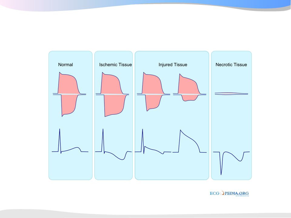
ZONE OF ISCHEMIA ZONE OF INJURY ZONE OF INFARCTION ISCHEMIA CAUSES INVERSION OF T WAVE DUE TO ALTERED REPOLARIZATION MUSCLE INJURY CAUSES ELEVATION OF S-T SEGMENT DURING RECOVERY (SUBACUTE AND CHRONIC STAGES) S-T SEGMENT OFTEN IS FIRST TO RETURN TO NORMAL, THEN T WAVE, DUE TO DISAPPEARANCE OF ZONES OF INJURY AND ISCHEMIA Q S DEATH (INFARCTION) OF MUSCLE CAUSES Q OR QS WAVES DUE TO ABSENCE OF DEPOLARIZATION CURRENT FROM DEAD TISSUE AND OPPOSING CURRENTS FROM OTHER PARTS OF HEART
 S-T SEGMENT OFTEN IS FIRST TO RETURN TO NORMAL, THEN T WAVE, DUE TO DISAPPEARANCE OF ZONES OF INJURY AND ISCHEMIA. Q. S. DEATH (INFARCTION) OF MUSCLE CAUSES Q OR QS WAVES DUE TO ABSENCE OF DEPOLARIZATION CURRENT FROM DEAD TISSUE AND OPPOSING CURRENTS FROM OTHER PARTS OF HEART.")
47
Anterolateraal infarct

48
ECG patterns and LAD ischemia
AVR AVL I II AVF III V5 AVF AVR V6 AVL V3 V4 V2 V1 This is from the TIMI IIIb study and troponin I Proximal LAD occlusion. Global ischemia of the whole anterior and septal aspect of the left ventricle. The ST segment vector points in a superior direction, because the anterobasal segment is the dominant ischemic area. Related ECG changes. The superiorly oriented ST vector leads to ST changes, such as ST elevation in lead AVR and V1 with reciprocal ST depression in the inferior leads and in leads V5 and V6. Adapted from Wellens et al.

49
ECG patterns and LAD ischemia
AVR AVL I II AVF III V1 V3 V4 V2 V5 AVF AVR V6 This is from the TIMI IIIb study and troponin I Related ECG changes. The inferiorly directed ST vector leads to ST depression in lead AVR, and ST segment elevation in the inferior leads. Distal LAD occlusion. The ST vector points more inferiorly due to ischemic dominance of the inferoapical area. Adapted from Wellens et al.

50
ECG patterns and LAD ischemia
AVR AVL I II AVF III This is from the TIMI IIIb study and troponin I Perfusion areas of the left anterior descending branch of the left coronary artery (LAD) Myocardial areas perfused by branches from the LAD include the septal, lateral and inferoapical area. The ST segment elevation vector is the result of the amount of ischemia in these respective areas. Possible occlusion sites related to the main branches of the LAD Upper left. Proximal to first septal and first diagonal branch. Lower left. Distal to first septal, proximal to first diagonal branch. Upper right. Distal to first diagonal, proximal to first septal branch. Lower right. Distal to both branches. Adapted from Wellens et al.
 Myocardial areas perfused by branches from the LAD include the septal, lateral and inferoapical area. The ST segment elevation vector is the result of the amount of ischemia in these respective areas. Possible occlusion sites related to the main branches of the LAD Upper left. Proximal to first septal and first diagonal branch. Lower left. Distal to first septal, proximal to first diagonal branch. Upper right. Distal to first diagonal, proximal to first septal branch. Lower right. Distal to both branches. Adapted from Wellens et al.")
51
ECG patterns and LAD ischemia
ECG criteria to identify site of occlusion in the LAD (From: Engelen et al. (49) Criterium Occlusion site Sens Spec PPA NPA RBBB Proximal to S1 14 100 62 ST V1 > 2.5mm 12 61 ST AVR 43 95 86 70 ST V5 17 98 88 Q AVL Proximal to D1 44 85 67 69 ST II ≥ 1.0mm Proximal to S1/D1 34 93 68 Q V5 Distal to S1 24 71 53 ST AVL Distal to D1 22 87 46 No ST III Distal to S1/D1 41 92 This is from the TIMI IIIb study and troponin I NPA = negative predictive accuracy PPA = positive predictive accuracy RBBB = Right bundle branch block Sens = Sensitivity Spec = Specify
 Criterium. Occlusion site. Sens. Spec. PPA. NPA. RBBB. Proximal to S ST V1 > 2.5mm ST AVR ST V Q AVL. Proximal to D ST II ≥ 1.0mm. Proximal to S1/D Q V5. Distal to S ST AVL. Distal to D No ST III. Distal to S1/D This is from the TIMI IIIb study and troponin I. NPA = negative predictive accuracy PPA = positive predictive accuracy. RBBB = Right bundle branch block Sens = Sensitivity Spec = Specify.")
52
Tombstoning LAD

53
♂ 73 jr. A: Bij presentatie 90 min AP VG: AF
ST-elevaties in I, aVL, aVR, V1-5, LAD (proximaal; ST depressies II, III, aVF, V6 voor septale en L-as, 1e grd AVB, RBTB, LAHB diagonale tak) / HS
 / HS.")
54
Bloedvoorziening geleidingssysteem
LAD CX RBB His AF PF AV node RCA SA knoop: RCA in 55% AV knoop: RCA in 90% (RCX) Bundel van His: RCA / LAD Rechter bundeltak: LAD (S1) Linkerbundeltak: Anticus: LAD Posticus: LAD/ RCA
 Bundel van His: RCA / LAD. Rechter bundeltak: LAD (S1) Linkerbundeltak: Anticus: LAD. Posticus: LAD/ RCA.")
55
Geleidingsproblemen bij AMI
Sinus bradycardie/ -arrest -/ totaal AV Blok(RCA): Vagaal/ ischemisch/ neurolgisch (Bezold- Jarisch reflex), humoraal (pH, adenosine) atropine/ aminophylline 10-20%, (meestal) smalcomplex escape ritmeHR: 40-60/ min, voorbijgaand, wel mortaliteit (2-3x) Totaal AV blok (LCA): Ischemisch (septale necrose) 5%, breedcomplex escape ritmeHR: <40/ min, voorbijgaand, wel mortaliteit (4x), pacemakerindicatie (primaire PCI) Bij proximale LAD: RBTB, LAHB, 1e grds AVB
: Vagaal/ ischemisch/ neurolgisch (Bezold- Jarisch reflex), humoraal (pH, adenosine) atropine/ aminophylline %, (meestal) smalcomplex escape ritmeHR: 40-60/ min, voorbijgaand, wel mortaliteit (2-3x) Totaal AV blok (LCA): Ischemisch (septale necrose) 5%, breedcomplex escape ritmeHR: <40/ min, voorbijgaand, wel mortaliteit (4x), pacemakerindicatie (primaire PCI) Bij proximale LAD: RBTB, LAHB, 1e grds AVB.")
56
LVH

57
LBTB

58
Infarct bij LBTB Concordantie ipv discordantie
Toename discordantie in V1 (>5 mm)
")
59
Hyperkaliemie

60
Pericarditis

61
Quiz

Verwante presentaties

 SEPSIS>")


